Common Types of Moles
Congenital Moles
Congenital moles, also called congenital nevi, appear at birth or within the first year of a baby’s life. These moles are caused by melanocyte cells in the epidermis (outer layer of skin), the dermis (middle layer of skin), or both. They are frequently called birthmarks and come in all sizes. Congenital moles could possibly develop into melanoma as you age and should be monitored by a skilled Gainesville dermatologist.
Atypical Moles
Atypical moles, or dysplastic nevi, are irregular moles displaying irregular symptoms including blurry borders, varying color, increased size, and flat/raised characteristics. Dysplastic nevi exhibit similarities to precancerous and cancerous moles, but most are harmless. When a person has multiple atypical moles, however, they have an increased risk of skin cancer and should perform regular checks to document changes.
Intradermal Nevi
Intradermal moles are the color of skin that blends in with the rest of the skin around them. They are located in the dermis, or the middle layer of skin, which explains why they aren’t as dark a junctional melanocytic moles. These moles are very common and usually benign.
Acquired Moles
Unlike congenital moles, acquired moles show up during later childhood and adulthood and are the most common type of mole. In general, acquired moles pose no risk and are benign, though sometimes they can develop into cancerous moles as the person ages.
Compound Nevi
Compound nevi combine features from junctional and intradermal moles, with melanocytes located in both the epidermis-dermis intersection and the dermis. Like junctional moles, they are slightly raised while flat on the edges and have distinct borders around them.
Halo Nevi
Halo moles are still a mystery to many doctors — they have a ring of skin around them that’s lost all pigmentation due to inflammatory cells infiltrating. The reason for this still puzzles doctors, but these moles are safe and require no treatment outside of cosmetic reasons.
Junctional Melanocytic Nevi
Junctional melanocytic moles occur at the meeting point of the dermis and epidermis after melanocyte cells cluster. These moles are somewhat raised with dark pigmentation and defined borders. It’s common for them to show up as people age since melanocytes burrow into deeper layers of skin later in life.
Inspecting Moles
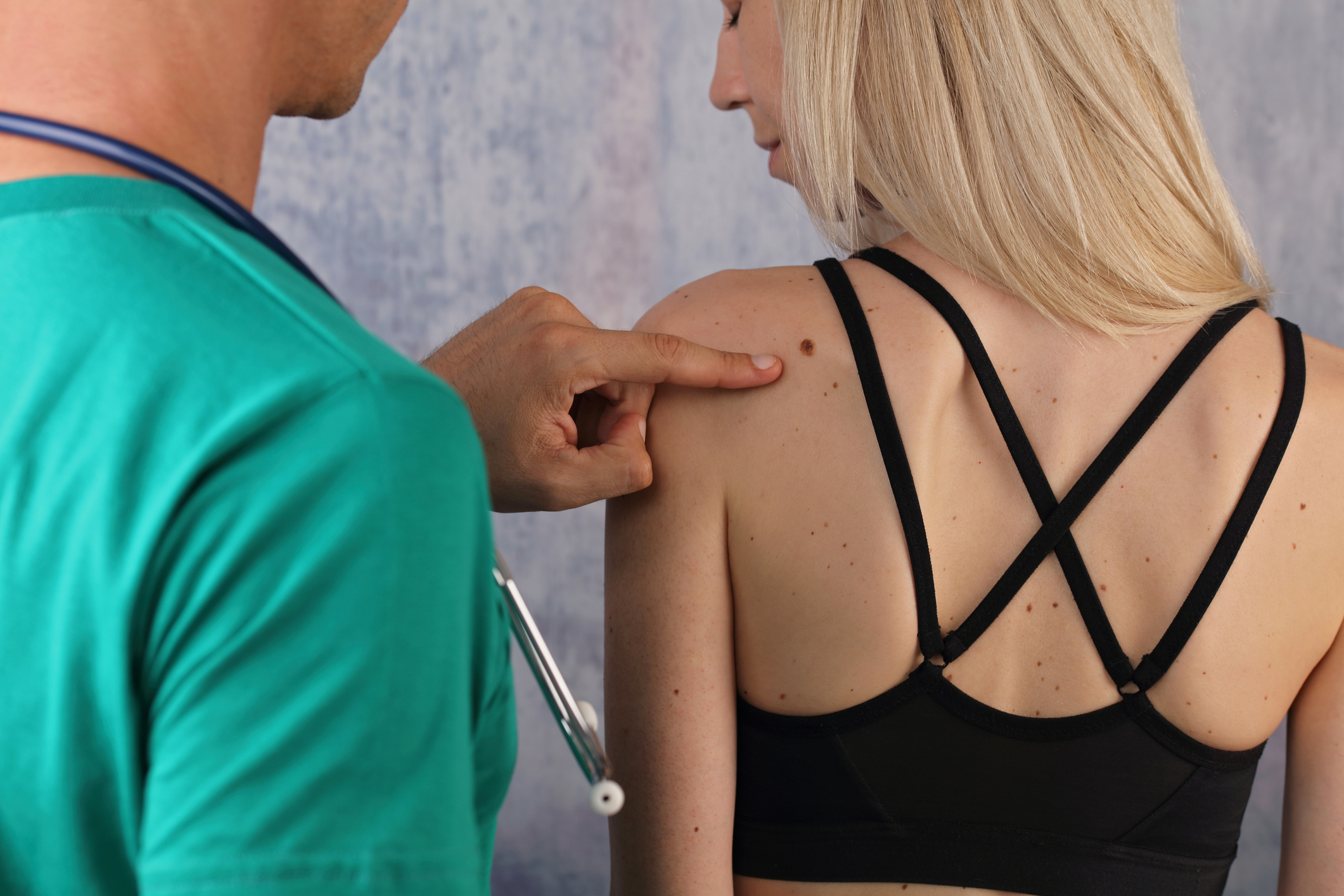
It’s important to frequently check on atypical moles and cite abnormal changes in size, color, shape, etc. The best way to examine moles on the body is with mirrors, but having someone help can also be useful if the moles are in a hard-to-see area. You should check the moles at least once a month and remove all nail polish from fingernails and toenails, as they can be sites of possible skin cancer. It’s vital to look at all areas of the skin including the scalp, hands, feet, genitals, and back.
Identifying Melanoma From Moles
Usually moles are harmless; however, the appearance of many atypical moles or moles that have changed in color could be early signs of melanoma, a type of skin cancer. The ABCs can help identify this type of skin cancer.
- Asymmetry: One side of the mole looks different from the other side. This is the time to look for irregular shapes and edges.
- Borders: The borders are jagged and indistinct, and pigmentation is spread outside the mole’s outline.
- Color: Inspect for growths that have changed colors, have multiple colors, or uneven colors. Different amounts of black, white, red, grey, blue, brown, or tan may be seen.
- Diameter: The size of the mole increases to larger than a quarter-inch, or a pencil head eraser. While some melanomas can be tiny, most are bigger than a ¼ inch.
- Evolution: Keep an eye out for moles that change in size, color, elevation or shape, especially if part of the moles turns black. Irritation, itching or bleeding may occur and requires attention from a skilled Gainesville dermatologist.
Diagnosing Melanoma
In addition to the ABCs, dermatologists will use a magnifying tool called a dermatoscope to help distinguish between normal and atypical moles. If the moles show any of these signs or are a cause for concern, the best way to be diagnosed is to remove a sample of tissue from the growth and have it examined by a professional at Gainesville Dermatology and Skin Surgery.
What Happens If The Mole Is Precancerous Or Cancerous?
If the mole is precancerous or cancerous, the next step is to have a complete skin exam and a thorough physical, concentrating on the lymph nodes where melanoma usually begins to spread. Dermatologists will use this information to determine the stage and exact location of melanoma before moving on to treatment options.
Melanoma treatment most often involves surgery, since the goal is to remove all the cancerous areas. If found early, this may be the only treatment needed. However, melanoma spreads quickly, so a combination of surgery and medication is most likely.
Melanoma Prevention
There are a few ways to help limit the growth of moles and chance at developing melanoma.
- Avoid peak sun times. For Gainesville residents, the sun’s ultraviolet rays are strongest between 10 a.m. and 3 p.m., so schedule activities for other times in the day, even when it’s cloudy.
- Use sunscreen in all seasons. Always apply sunscreen 30 minutes before going outside with a recommended SPF of 15 or higher.
- Cover up. Wear clothing that can shield your skin from UV rays including sunglasses, sunhats, long sleeves, and UV-ray protective clothing.
- Avoid tanning beds and lamps. These both emit UV rays and can increase your risk of getting skin cancer.
If you have questions or want to set up an appointment to examine your moles, contact us today. Early detection is imperative and can increase your chances of successful treatment of melanoma or other skin cancers.

